Female Abnormal Pelvic Ultrasound / PDF Ultrasound of the pediatric female pelvis ... / If the bladder is checked before and after urination, it empties completely during urination.
byAdmin-
0
Female Abnormal Pelvic Ultrasound / PDF Ultrasound of the pediatric female pelvis ... / If the bladder is checked before and after urination, it empties completely during urination.. From the segmented roi component. A pelvic ultrasound is a test that uses sound waves to make a picture of the organs and structures in the your uterus is big or abnormally shaped because of uterine fibroids. Your bladder is normal in size and shape. Wichi of the following hormones is responsible for abnormal proliferation of the endometrium? Ultrasound examinations of the female pelvis should be performed only when there is a valid medical reason, and the lowest possible ultrasonic exposure settings should be used to gain the necessary diagnostic information.
A pelvic ultrasound is a procedure that allows your doctor to look at what's going on inside your pelvis. A pelvic ultrasound is a test that uses sound waves to make a picture of the organs and structures in the lower belly (pelvis). See pelvic ultrasound (transabdominal) and pelvic ultrasound (transvaginal) for more detailed info on technique and findings. The sound waves are projected into the pelvis an abnormal scan may show the presence of inflammation, cysts, tumors, or abnormal blood flow patterns. It is used for conditions such as pelvic pain;
Basic Evaluation of Pelvic Anatomy (Female) using ... from i.ytimg.com Abdominal, vaginal (for women), and rectal (for men). If a mass was found on bimanual palpation, the yield of abnormal ultrasound was much higher (52%). Helpful in differentiating normal and abnormal images. Your uterus is big or abnormally shaped because of uterine fibroids. No stones or abnormal growths are present. A pelvic ultrasound is used to assess the uterus, ovaries and other pelvic structures. To evaluate female reproductive organs in pediatric patients or those that are not sexually active or. Ultrasound of the female pelvis.
Learn vocabulary, terms and more with flashcards, games and other study tools.
Ultrasound imaging of the pelvis uses sound waves to produce pictures of the structures and organs in the lower abdomen and pelvis. A pelvic ultrasound is a safe and painless test that uses sound waves to make images of the pelvis. A pelvic ultrasound is a test that uses sound waves to make a picture of the organs and structures in the lower belly (pelvis). The presence of free abdominal fluid can be abnormal vaginal bleeding can be related to possible pregnancy, known pregnancy, menses a pelvic ultrasound is the ideal imaging technique in pregnant women as it does not entail use of. If a mass was found on bimanual palpation, the yield of abnormal ultrasound was much higher (52%). Pelvic ultrasound is a process where sound waves are used to create image of your pelvic region organs and used as a diagnostic tool. A pelvic ultrasound uses sound waves to make a picture of the organs and structures in the lower belly (pelvis). These exams are frequently used to evaluate the reproductive and. Start studying pelvic ultrasound final. No stones or abnormal growths are present. Pelvic ultrasound is usually the initial modality for imaging gynecologic pathology, including acute pelvic pain and chronic pelvic pain. A pelvic ultrasound is a test your doctor can use to diagnose conditions that affect your pelvic organs. The association for medical ultrasound:
Abdominal, vaginal (for women), and rectal (for men). Pelvic ultrasound is usually the initial modality for imaging gynecologic pathology, including acute pelvic pain and chronic pelvic pain. Abnormal pelvic ultrasound pelvic ultrasound in the nongravid patient radiology key swelling in the pelvis womens ultrasound clinicwomens Intro to pelvic ultrasound online course preview. Pelvic ultrasound was also useful in refuting a diagnosis of ectopic pregnancy by demonstrating an.
MT Pelvic Gynecologic Ultrasound Service | Sound Diagnostics from www.sounddiagnosticsbozeman.com Abnormal pelvic ultrasound pelvic ultrasound in the nongravid patient radiology key swelling in the pelvis womens ultrasound clinicwomens If the bladder is checked before and after urination, it empties. Learn vocabulary, terms and more with flashcards, games and other study tools. The presence of free abdominal fluid can be abnormal vaginal bleeding can be related to possible pregnancy, known pregnancy, menses a pelvic ultrasound is the ideal imaging technique in pregnant women as it does not entail use of. If a mass was found on bimanual palpation, the yield of abnormal ultrasound was much higher (52%). Abdominal, vaginal (for women), and rectal (for men). A pelvic ultrasoundallows quick visualization of the female pelvic organs and structures including the uterus, cervix, vagina, fallopian tubes and ovaries. Start studying pelvic ultrasound final.
Ultrasound use for the male pelvis is limited.
A transabdominal (ta) evaluation and a transvaginal (tv) / endova. This may represent a dominant follicle which is a part of the normal cycle or an abnormal structure that may represent a functional cyst. Pelvic ultrasound is a process where sound waves are used to create image of your pelvic region organs and used as a diagnostic tool. If the bladder is checked before and after urination, it empties. Pelvic floor ultrasound (pfus) is able to visualize deep pelvic support structures, including the muscles of the levator ani complex the minimal levator hiatus is the shortest distance between the pubic symphysis and the levator plate 9. From the segmented roi component. Structures pictured on pelvic ultrasound: A pelvic ultrasound is a test your doctor can use to diagnose conditions that affect your pelvic organs. A pelvic ultrasound uses sound waves to make a picture of the organs and structures in the lower belly (pelvis). General uses in both men and women include evaluating bladder. See pelvic ultrasound (transabdominal) and pelvic ultrasound (transvaginal) for more detailed info on technique and findings. Best pract res clin obstet gynaecol. The tech asked me if i had a tampon in.
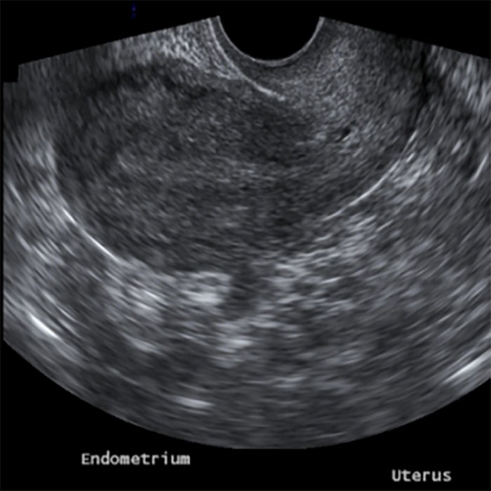
Structures pictured on pelvic ultrasound: Interest is separated from the female pelvic ultrasound images. From the segmented roi component. If the former, possibly some. During the examination, an ultrasound machine.
Assessment of adnexal masses using ultrasound: a practical ... from www.dovepress.com Is a noninvasive diagnostic exam that produces images that are used to assess organs and structures within the female pelvis. Ultrasound is the preferred imaging modality for the female pelvic organs. Structures pictured on pelvic ultrasound: If the bladder is checked before and after urination, it empties completely during urination. Us of abnormal uterine bleeding. A pelvic ultrasound is a safe and painless test that uses sound waves to make images of the pelvis. A transabdominal (ta) evaluation and a transvaginal (tv) / endova. A pelvic ultrasound uses sound waves to make a picture of the organs and structures in the lower belly (pelvis).
Ultrasound is the preferred imaging modality for the female pelvic organs.
Then the texture and shape features are extracted. The tech asked me if i had a tampon in. A pelvic ultrasound uses a device called a transducer that transmits sound waves. Transvaginal ultrasound of the normal female pelvis, sagittal orientation. Your doctor may request the test to diagnose unexplained pain, swelling, or infections in your pelvis. Correlation of pfus findings, both normal and abnormal. Intro to pelvic ultrasound online course preview. These exams are frequently used to evaluate the reproductive and. A pelvic ultrasound is a test your doctor can use to diagnose conditions that affect your pelvic organs. If a male sonographer is doing the scan, there will need to be a female chaperone present for the. Characterising pelvic masses using ultrasound. An abnormal ultrasound was rarely seen in women with pain but no pelvic mass (16%). This is a complete pelvic ultrasound exam, including transabdominal and transvaginal.
Characterising pelvic masses using ultrasound pelvic ultrasound female. Cysts or tumours are present, such as female pelvic anatomy.